Patient Resources
Your Spine/Back
Anatomy of the Spine
 The normal anatomy of the spine is usually described
by dividing up the spine into three major sections: the
cervical, the thoracic and the lumbar spine. Below the
lumbar spine is a bone called the sacrum, which is part
of the pelvis.
The normal anatomy of the spine is usually described
by dividing up the spine into three major sections: the
cervical, the thoracic and the lumbar spine. Below the
lumbar spine is a bone called the sacrum, which is part
of the pelvis.
Each section is made up of individual bones called
vertebrae. There are seven cervical vertebrae, 12
thoracic vertebrae, and five lumbar vertebrae.
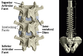
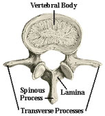
An individual vertebra is made up of several parts.
The body of the vertebra is the primary area of weight
bearing and provides a resting place for the fibrous discs
that separate each of the vertebrae. The lamina covers the spinal canal, the large hole in the center of the
vertebra through which the spinal nerves pass. The spinous process is the bone you can feel when running
your hands down your back. The paired transverse processes are oriented 90 degrees to the spinous process
and provide attachment for back muscles.
 There are four facet joints associated with each vertebra. A pair that face upward
and another pair that face downward. These interlock with the adjacent vertebrae
and provide stability to the spine.
There are four facet joints associated with each vertebra. A pair that face upward
and another pair that face downward. These interlock with the adjacent vertebrae
and provide stability to the spine.
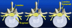
The vertebrae are separated by intervertebral discs that act as cushions between
the bones. Each disc is made up of two parts. The hard, tough outer layer called
the annulus surrounds a mushy, moist center termed the nucleus. When a disc
herniates or ruptures, the soft nucleus spurts out through a tear in the annulus,
and can compress a nerve root. The nucleus can squirt out on either side of
the disc or in some cases both sides. The amount of pain associated with a
disc rupture often depends upon the amount of nucleus that breaks through
the annulus, and whether it compresses a nerve. To help alleviate the pain, a
laminotomy/microdiscetomy may be performed.
Herniated discs
 To alleviate the pain of a ruptured or herniated intervetebral
disc (see Spinal Anatomy), a laminotomy/microdiscetomy
may be performed. This surgical procedure is carried out in
two steps beginning with the laminotomy. Lamina is the Latin
name given to the bone protecting the spinal canal, and otomy
means opening or hole. The laminotomy simply opens up the
spinal canal in order to visualize the pinched nerve root.
To alleviate the pain of a ruptured or herniated intervetebral
disc (see Spinal Anatomy), a laminotomy/microdiscetomy
may be performed. This surgical procedure is carried out in
two steps beginning with the laminotomy. Lamina is the Latin
name given to the bone protecting the spinal canal, and otomy
means opening or hole. The laminotomy simply opens up the
spinal canal in order to visualize the pinched nerve root.
Once this is accomplished, the second procedure, the microdiscetomy, is performed. A high powered
stereoscopic microscope is used to provide illumination and magnification to allow the nerve and surrounding
structures to be visualized clearly through an incision less than one inch long. The nerve root is carefully
protected with a specialized retractor, and protruding disc fragments, along with any remaining loose or
degenerated disc material, are then removed. The small hole left in the annulus will regenerate in four to six
weeks and fill in with new disc material.
Osteoporosis
Detection with Bone Mineral Density Testing
There has been much recognition of osteoporosis as a women's health problem, but five million American
men are also affected. Even teenagers are not immune, particularly girls. Osteoporosis causes the bones
to weaken and fracture under the slightest stress. If detected early, it can be prevented and treated before
significant bone loss occurs. Bone tissue can be affected by age, heredity, unhealthy habits, diet, sex
hormones, physical inactivity and certain medications.
Osteoporosis can be a complication of any chronic disease involving the lungs, liver, kidneys, GI tract,
hormones, and rheumatoid arthritis. Bone loss can occur as a result of long-term use of steroids, thyroid
hormone, some anti-convulsant drugs and chemotherapies.
Low levels of sex hormones are the major cause of osteoporosis. Men with low testosterone levels can be
treated with replacement therapy.
Women who complete menopause or suffer loss of menstrual periods should investigate the possibility of
bone loss with a BMD test. The BMD test is quick, painless, similar to x-ray, and can predict fracture risk.
Unhealthy habits such as smoking and excessive alcohol intake are known risk factors. A regular regimen of
improved physical activity, especially weight-bearing exercise or the use of resistance machines, can prevent
or slow the inevitable loss of bone with aging.
It is important to tell your doctor if you detect any loss of height, have sudden back pain or suffer a fracture
with little trauma. A medical workup would include a complete medical history, x-rays and blood and urine test.
Your doctor can order a bone mineral density (BMD) test at OSC to detect low bone density.
Men and women who have risk factors should take calcium and, in some instances, Vitamin D, as a
preventative measure. Additional treatments for osteoporosis now include calcitonin, which comes in a nasal
spray, and bisphosphonate alendronate (Fosamax). Estrogen is the first line of defense against osteoporosis
and the decision whether or not to take estrogen should be carefully considered with your doctor, who can
recommend a specific program of treatment.
Scoliosis
 What is Scoliosis? And How is it Treated?
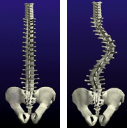
Everyone's spine has curves. These curves produce the normal
rounding of the shoulder and the sway of the lower back.
A spine with scoliosis has abnormal curves with a rotational deformity.
This means that the spine turns on its axis like a corkscrew. Compare
the more subtle curve of the normal spine to the severe curvature of the
scoliotic spine.
Scoliosis is a curvature of the spine that may have its onset in infancy
but is most frequently seen in adolescence. It is more common in
females by a 2:1 ratio. However, when curves in excess of 30 degrees
are evaluated, females are more frequently affected by a ration of approximately 8-10:1.
The cause of the most common form of scoliosis — idiopathic scoliosis — is unknown, but there are certainly
hereditary factors that are present.
Scoliosis causes shoulder, trunk and waistline asymmetry. In mild forms, the condition may be barely noticed;
whereas in severe forms there is significant disfigurement, back pain and postural fatigue, and it may be
associated with heart failure. Fortunately the majority of scoliosis cases need only close follow-up to watch for
worsening of the curve. Some cases require more aggressive treatment, which could include surgery.
Orthopedic surgeons are best qualified to evaluate and treat deforming spinal conditions like scoliosis.
However, a good resource for further information is:
The National Scoliosis Foundation
5 Cabot Place
Stoughton, MA 02072
Phone: (617) 341-6333
Fax: (617) 341-8333
Email: scoliosis@aol.com
Non-Operative Treatment
The non-operative treatment of scoliosis involves observing the deformity with examinations and repeated
x-rays. Under certain circumstances, when spinal growth remains, a brace may be used in combination with
follow-up x-rays. Physical therapy exercises have not been shown to be effective treatment for scoliosis.
Why Surgery?
Surgical treatment of scoliosis may be indicated for any of the following reasons: To prevent further
progression of the curve, to control the curve when brace treatment has failed, to improve an undesired
cosmetic appearance or for reasons of discomfort or postural fatigue.
Surgical Treatment of Scoliosis
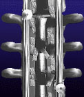 The most common surgical treatment for scoliosis is a spine fusion using special
stainless steel rods, hooks, and a bone graft. The rods are attached to the spine with
hooks and the curved portion of the spine is carefully straightened. Then, small strips
of bone graft are placed over the spine to fuse it in a straight position. As the bone
graft heals over the next several months, the spine becomes solid and will not curve
again. But the part of the spine that has not been fused will still be flexible, and allow
nearly normal overall movement.
The most common surgical treatment for scoliosis is a spine fusion using special
stainless steel rods, hooks, and a bone graft. The rods are attached to the spine with
hooks and the curved portion of the spine is carefully straightened. Then, small strips
of bone graft are placed over the spine to fuse it in a straight position. As the bone
graft heals over the next several months, the spine becomes solid and will not curve
again. But the part of the spine that has not been fused will still be flexible, and allow
nearly normal overall movement.
|
For Our Patients
Existing patients may utilize these convenient links to access forms, educational materials and manage their accounts.
Medical Questionnaire
Patient Forms
Convenient, Online
Patient Forms Are
Coming Soon!
Pay My Bill
INSURANCE PROVIDERS WE PARTICIPATE WITH:
Commercial
Blue Cross/Blue Shield
DMBA
HMA
HMAA
HMSA PPO & HMSA HMO
MDX
UHA
Government
Medicare
Medicaid
OHANA/Wellcare
Triwest/Tricare
UHC- Medicare Advantage
Other
Workers Compensation
No-Fault
|